Fungus Species in the Christopher B. Smith Preserve
Fungus Classification: Members of the Fungus Kingdom include yeasts, molds, mushrooms, puffballs, and bracket fungus. Some species are microscopic, while other are macroscopic. In this website, only the macroscopic fungi are discussed. All macrofungi produce sexual spores, created from the combination of genetic materials from two parents. The spores are produced and dispersed by fruiting bodies. The type of fruiting body determines whether it is classified as a member of Phylum/Division Ascomycota or Phylum/Division Basidiomycota. An ascomycete, also called a sac fungus, produces spores internally in a sac, called an ascus. This is the largest fungus phylum/division with over 64,000 species. A basidiomycete, also called a club fungus, produces sexual spores externally on the end of specialized cells called basidia.
Interactions in the Smith Preserve: Fungus species living in the Smith Preserve perform a variety of very important functions. Many are decomposers of organic matter and recycle nutrients. Some are parasitic. Some combine with algae and cyanobacteria to create lichens. Others live independently and provide food and habitat for other organisms. Scientists estimate there are 1.5 million species of fungus in the world, of which 100,000 species have been formally described. One can only imagine the number of different species living in the Smith Preserve.
Phylum/Division |
Class |
Order |
Family |
Species Name |
Common Name |
Ascomycota |
Dothideomycetes |
Capnodiales |
Capnodiaceae |
Capnodium sp. |
|
Ascomycota |
Laboulbeniomycetes |
Laboulbeniales |
Laboulbeniaceae |
Hesperomyces virescens |
|
Ascomycota |
Leotiomyetes |
Erysiphales |
Erysiphaceae |
Unknown |
|
Ascomycota |
Unknown |
Unknown |
Unknown |
Unknown |
|
Ascomycota |
Unknown |
Unknown |
Unknown |
Unknown |
|
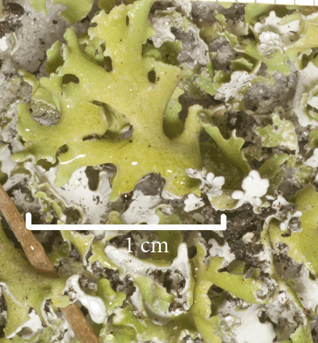
Ascomycota |
Ascomycetes |
Lecanorales |
Cladoniaceae |
Cladonia spp. |
|
Ascomycota |
Taphrinomycetes |
Taphrinales |
Taphrinaceae |
Taphrina caerulescens |
|
Basidiomycota |
Agaricomycetes |
Agaricales |
Lepiotaceae |
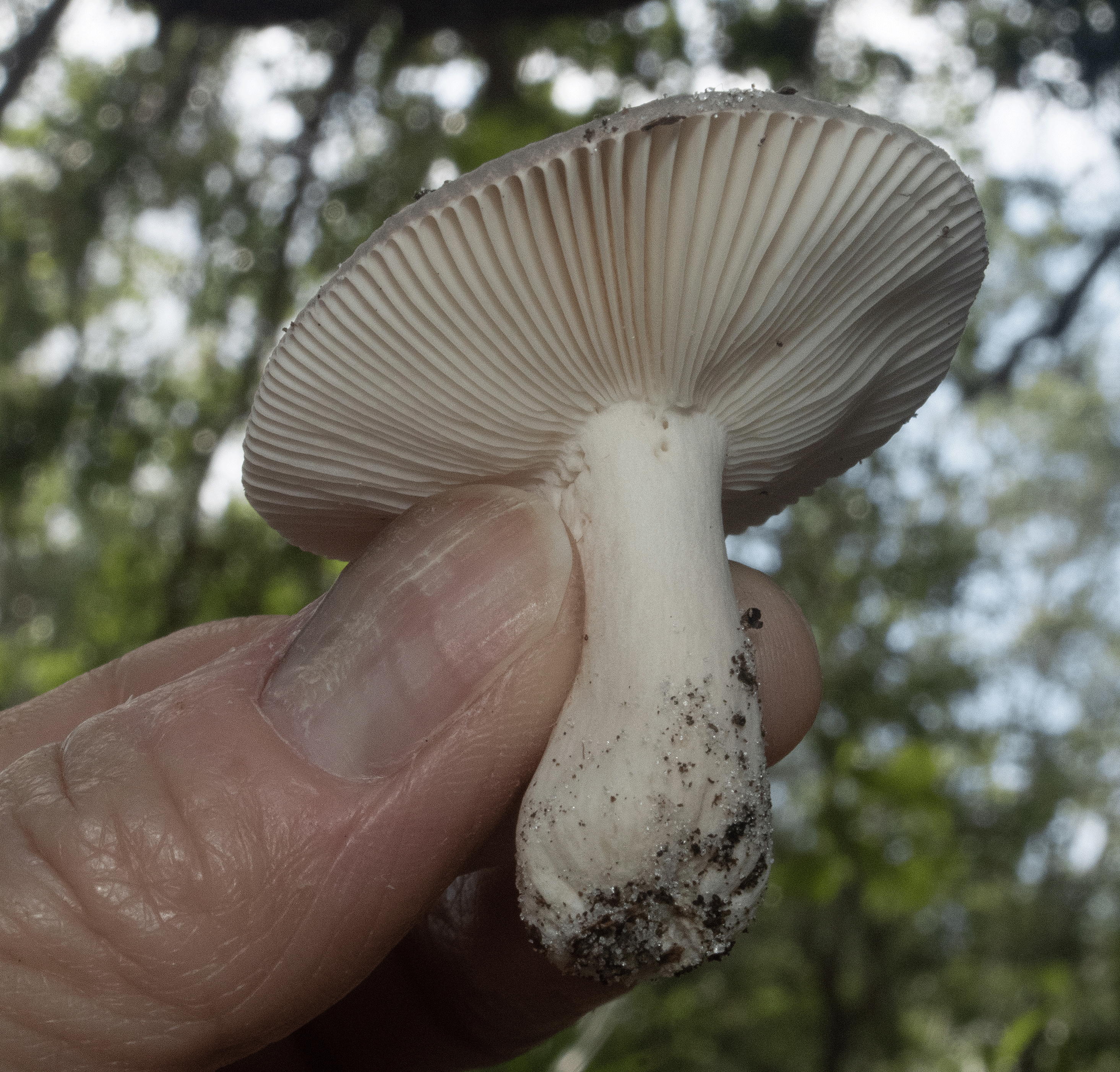
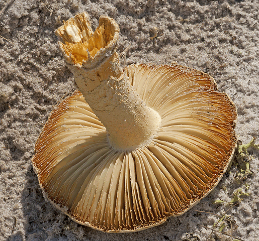
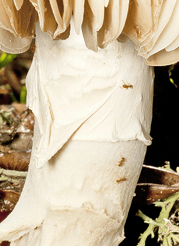
Unknown |
|
Basidiomycota |
Agaricomycetes |
Agaricales |
Lycoperdaeae |
Unknown |
|
Basidiomycota |
Agaricomycetes |
Agaricales |
Marasmiaceae |
Marasmiellus |
|
Basidiomycota |
Agaricomycetes |
Agaricales |
Schizophyllaceae |
Schizophyllum commune |
|
Basidiomycota |
Agaricomycetes |
Agaricales |
Unknown |
Unknown |
|
Basidiomycota |
Agaricomycetes |
Auriculariales |
Auriculariaceae |
Auricularia auricula-judae |
|
Basidiomycota |
Agaricomycetes |
Geastrales |
Geastraceae |
Geastrum sp. |
|
Basidiomycota |
Agaricomycetes |
Polyporales |
Ganadermataceae |
Ganaderma applanatum |
|
Basidiomycota |
Agaricomycetes |
Polyporales |
Ganodermataceae |
Ganoderma sp. |
|
Basidiomycota |
Agaricomycetes |
Polyporales |
Meruliaceae |
Gloeoporus taxicola ? |
|
Basidiomycota |
Agaricomycetes |
Polyporales |
Polyporaceae |
Hexagonia hydnoides |
|
Basidiomycota |
Agaricomycetes |
Polyporales |
Polyporaceae |
Lentinus sp. |
|
Basidiomycota |
Agaricomycetes |
Polyporales |
Polyporaceae |
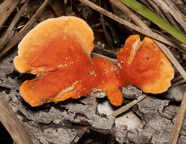
Pycnoporus sanguineus |
|
Basidiomycota |
Agaricomycetes |
Polyporales |
Polyporaceae |
Pycnoporus sp. |
|
Basidiomycota |
Agaricomycetes |
Polyporales |
Steccherinaceae |
Irpex lacteus |
|
Basidiomycota |
Agaricomycetes |
Hymenochaetales |
Hymenochaetaceae |
Hydnoporia olivacea |
|
Basidiomycota |
Agaricomycetes |
Unknown |
Unknown |
Unknown |
|
Basidiomycota |
Pucciniomycetes |
Septobasidiales |
Septobasidiaceae |
Septobasidium sp. |
|
Basidiomycota |
Agaricomycetes |
Unknown |
Unknown |
Unknown |
|
Basidiomycota |
Agaricomycetes |
Unknown |
Unknown |
Unknown |
|
Basidiomycota |
Agaricomycetes |
Unknown |
Unknown |
Unknown |
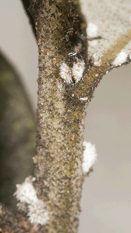
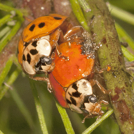
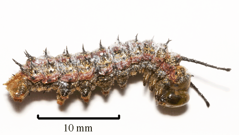
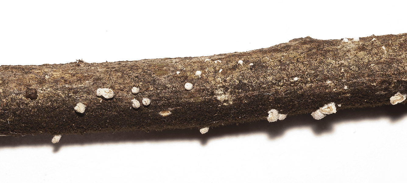
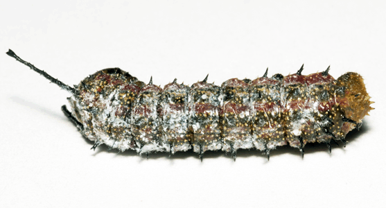 There are several fungal groups that can be entomopathogenic fungi. Since these fungi are considered natural agents of mortality and environmentally safe, there is interest in the use and manipulation of these fungi for biological control of pest insects and other arthropods.
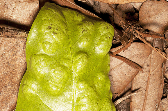
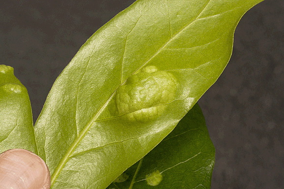
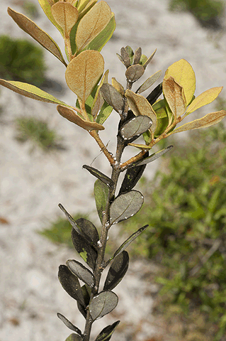
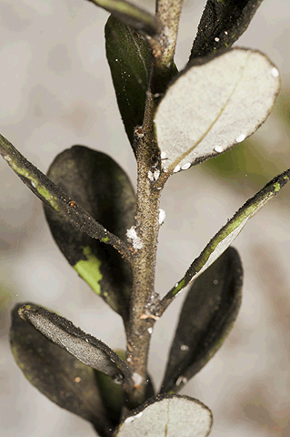
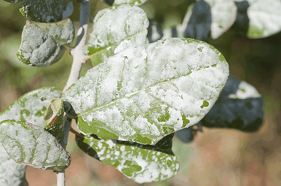
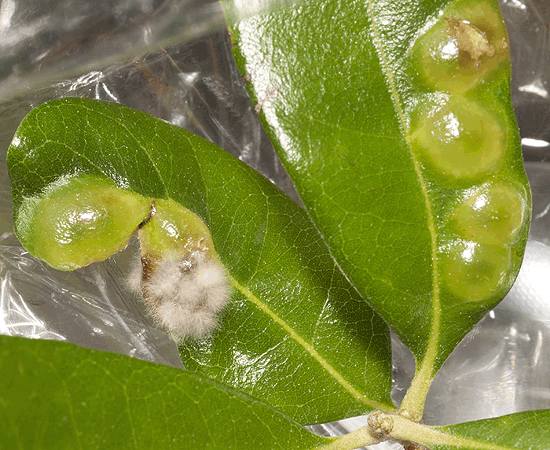 On February 28, 2017, the photographs at left and below were taken of another example of what appears as Taphrina caerulescens. In these photographs, the raised surfaces were on the top of the laurel oak leaves. The second image is an enlargement of the blister that appears to have hyphae.
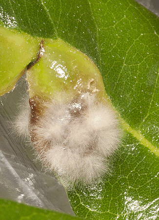


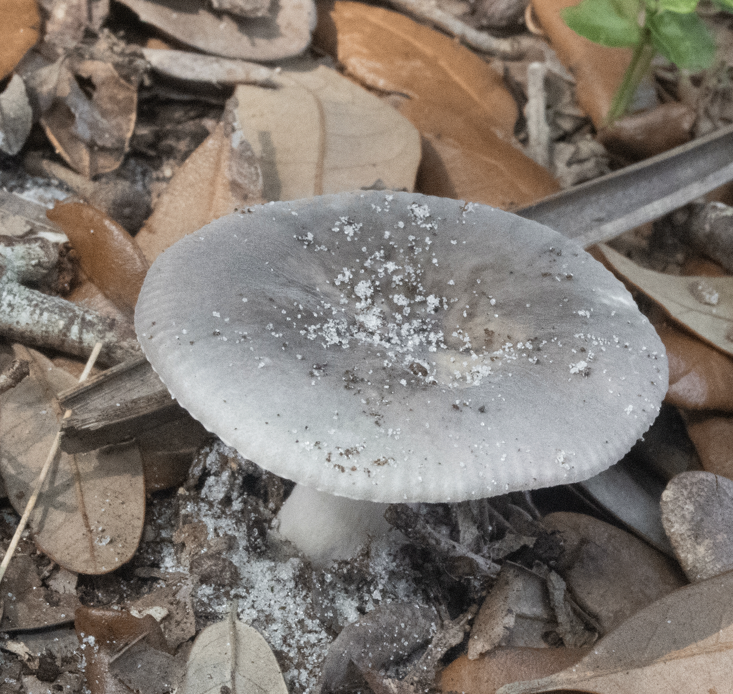
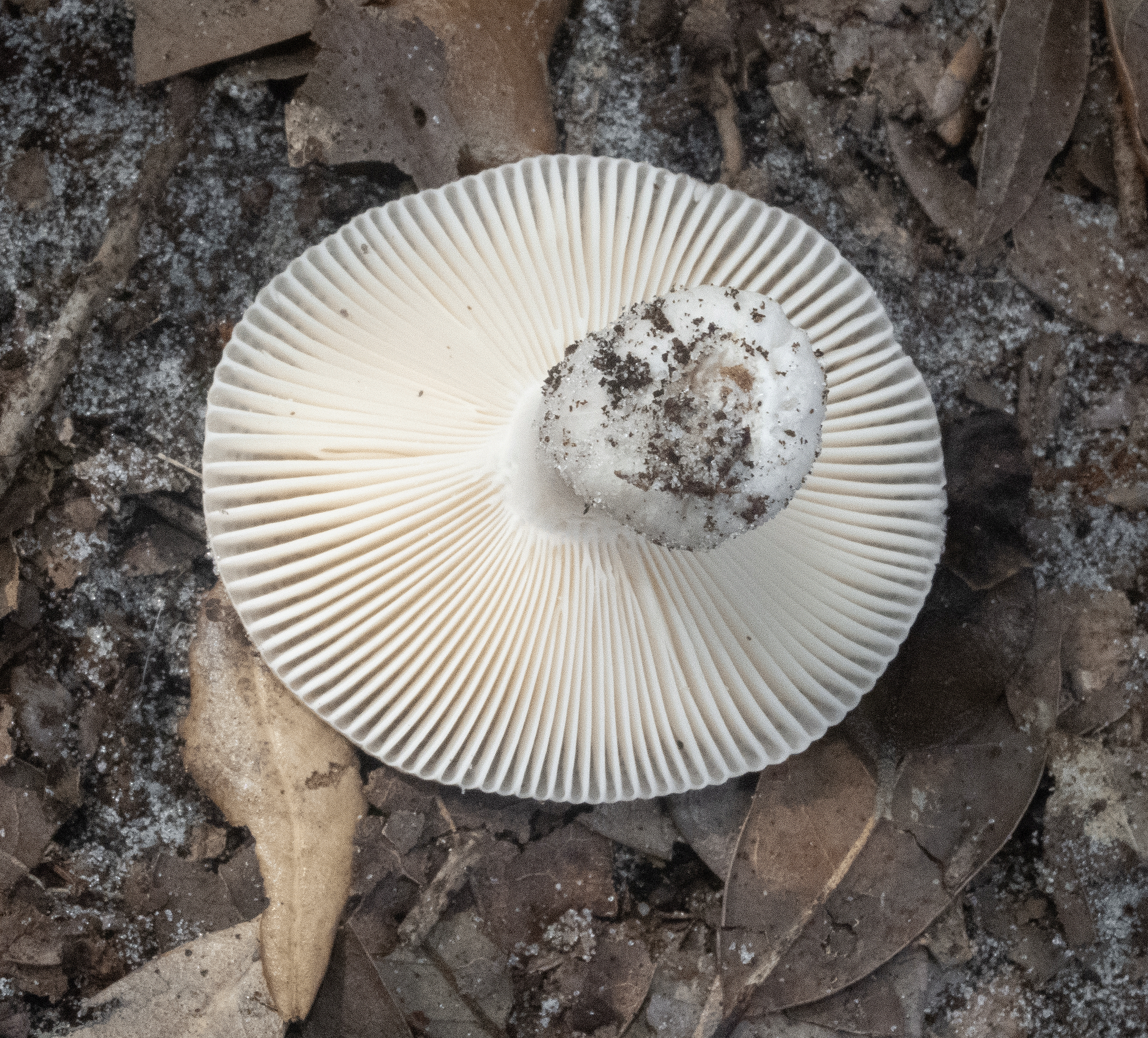
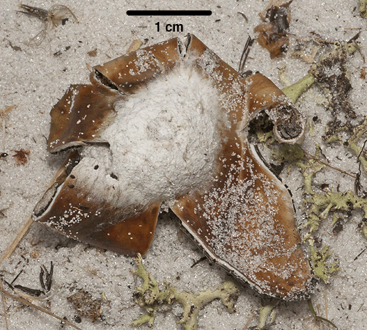
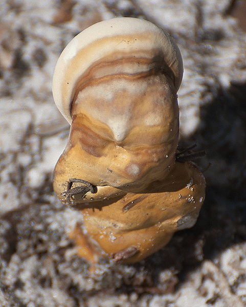
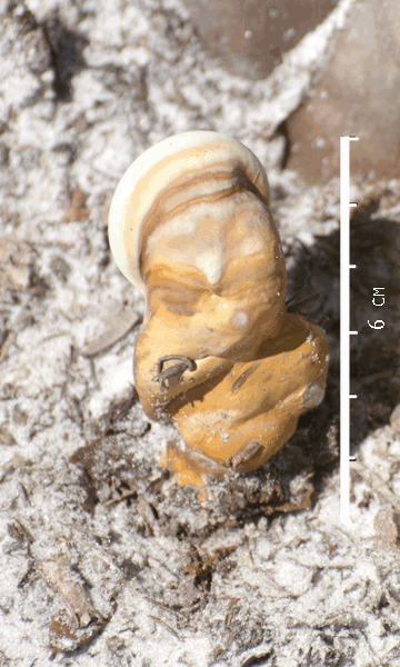
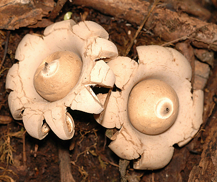 Geastrlum sp.
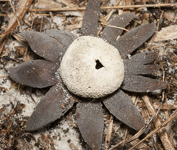
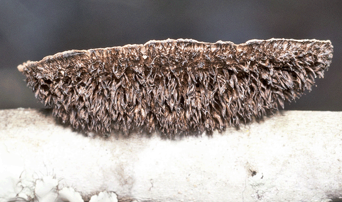
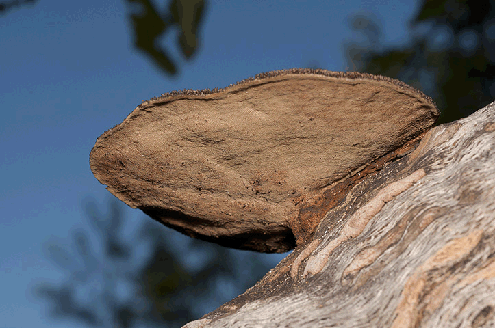
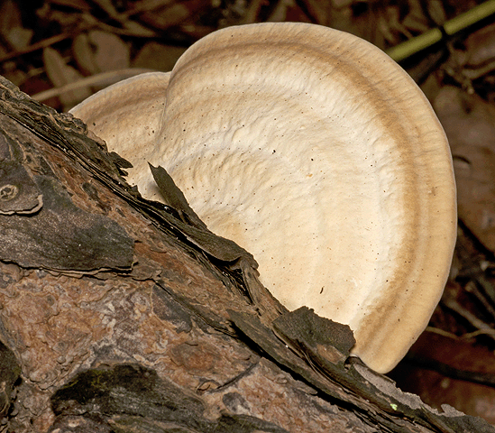 Artist's bracket is hard and shelf-like with gray-brown banding. The bottom is white and turns brown when scratched.
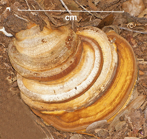
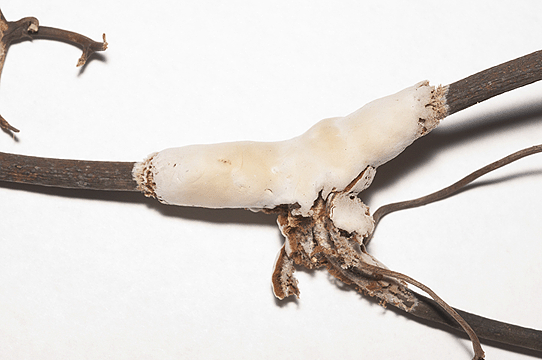
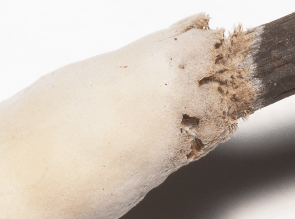
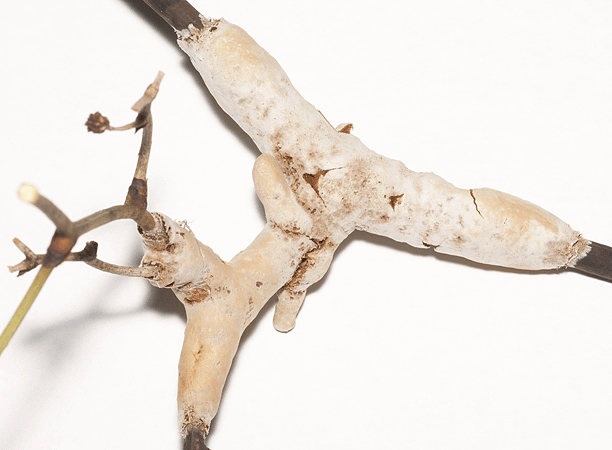
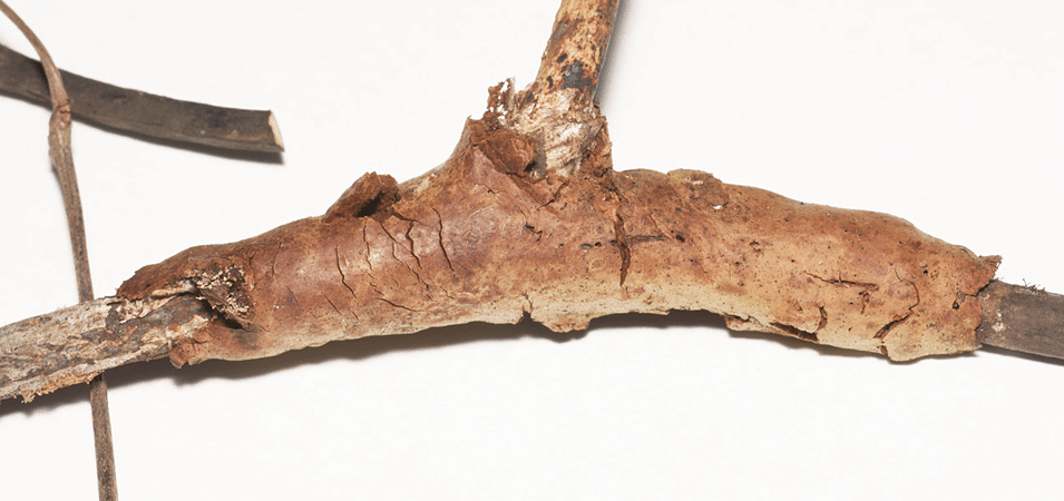
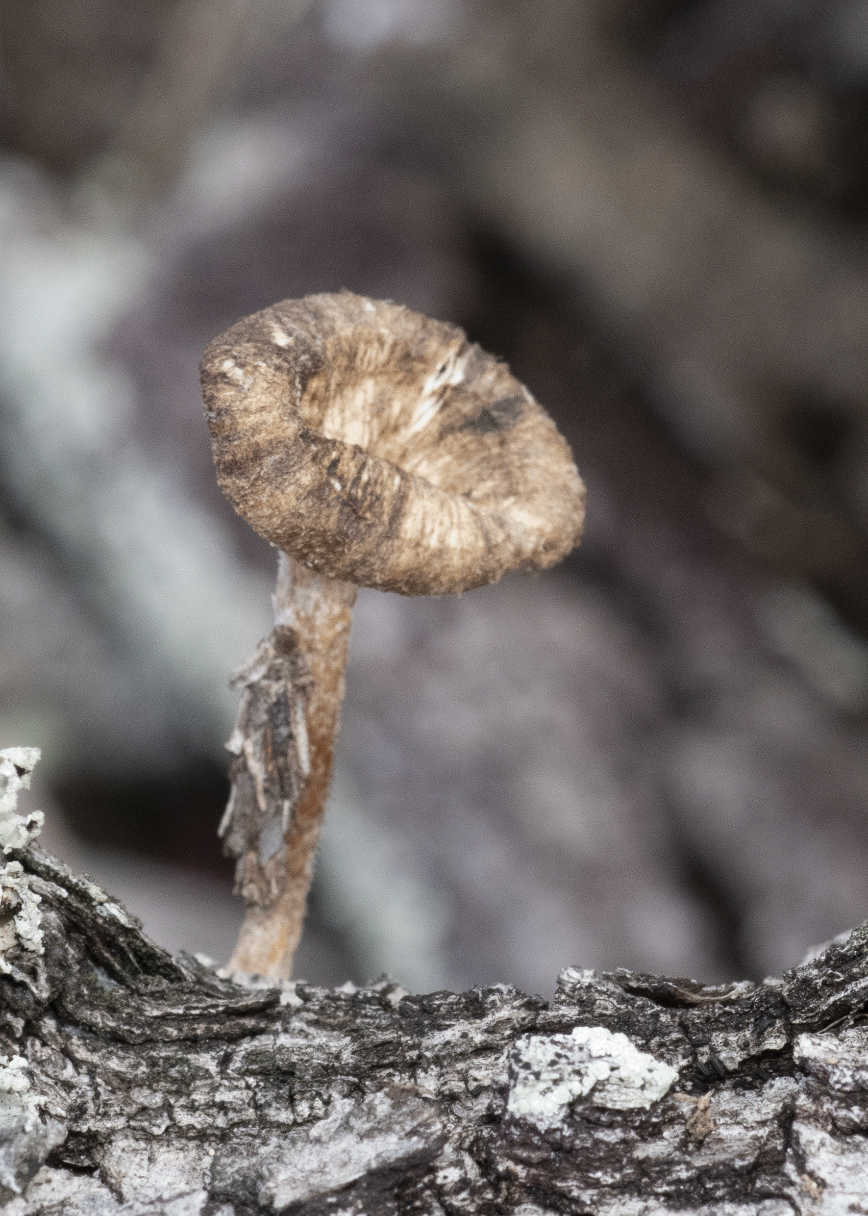
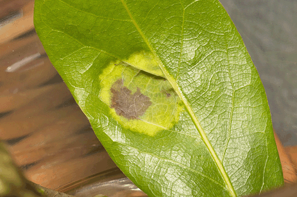
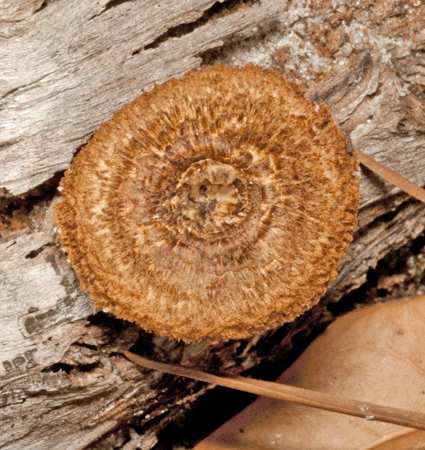
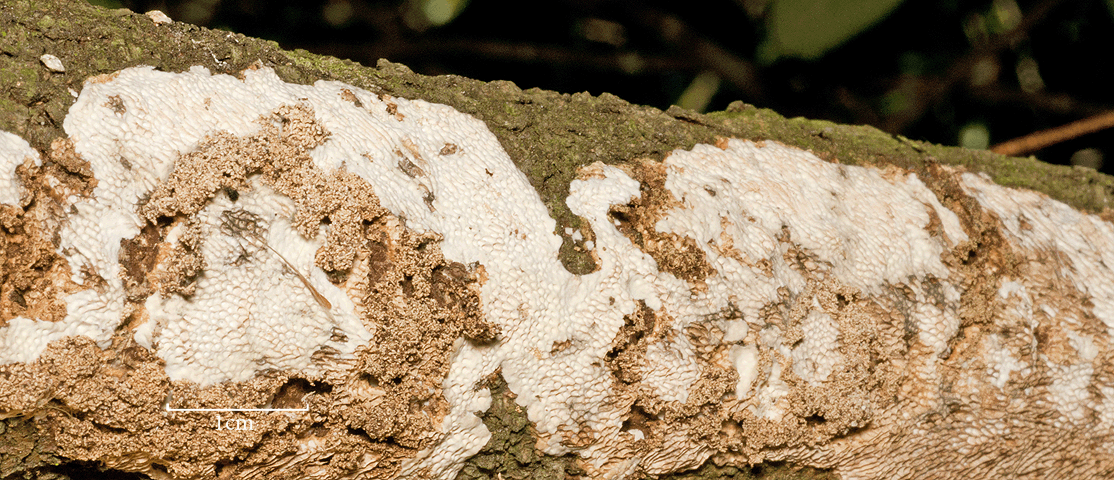
 This fungus does not have any nodular structures and it is rust-brown.
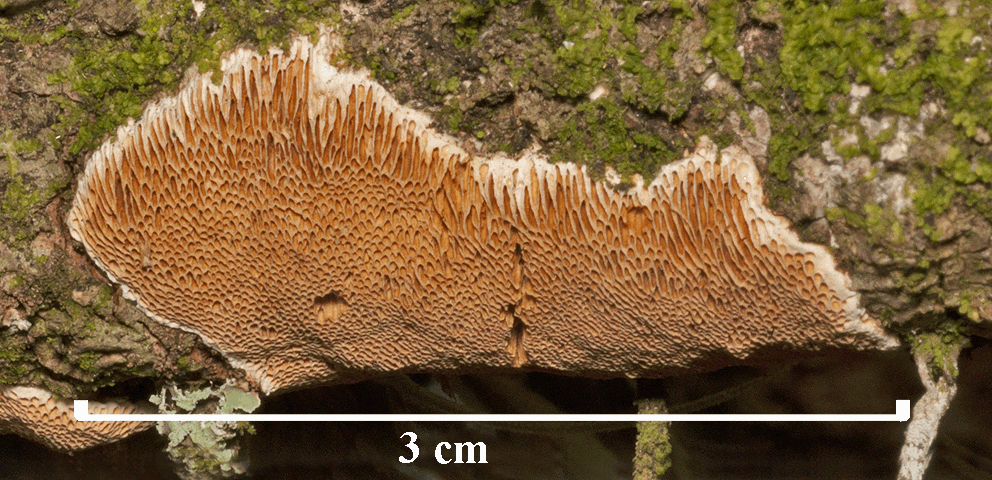
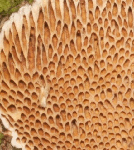
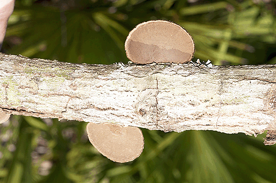
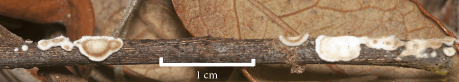
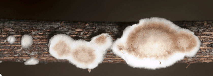
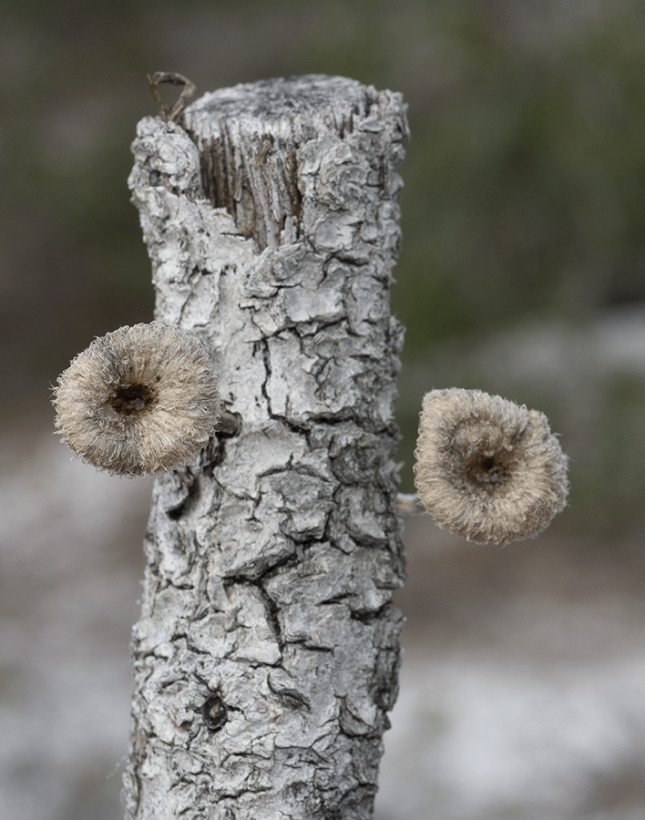
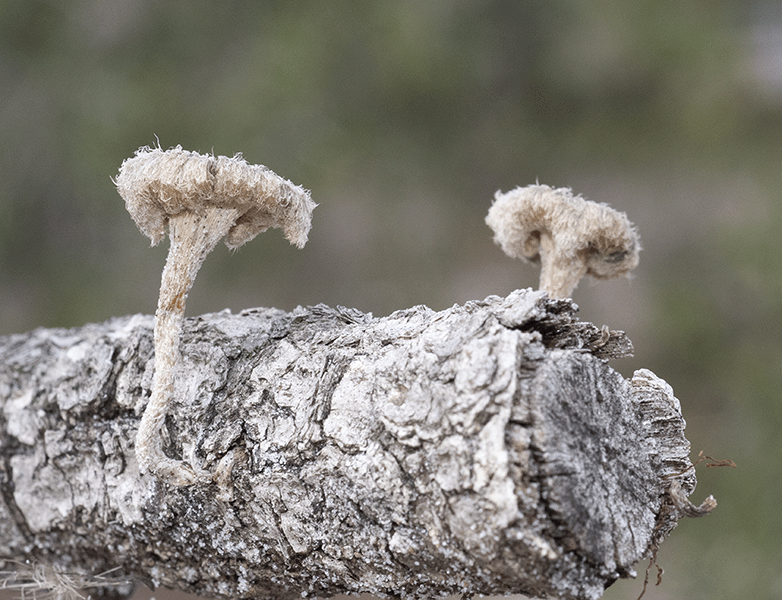
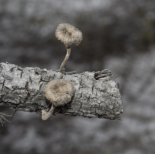
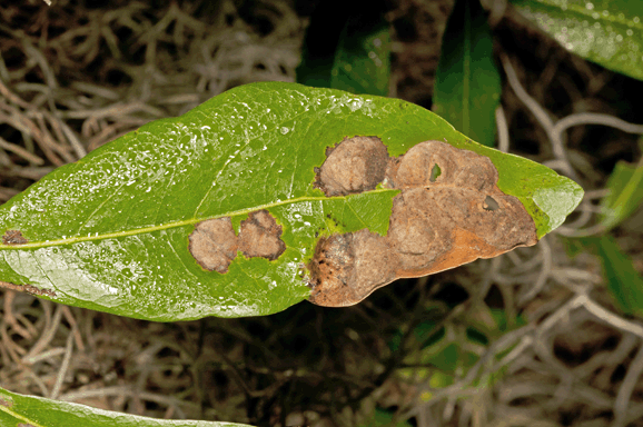
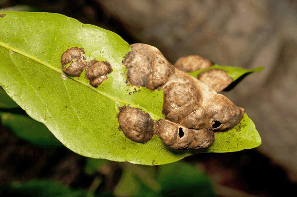
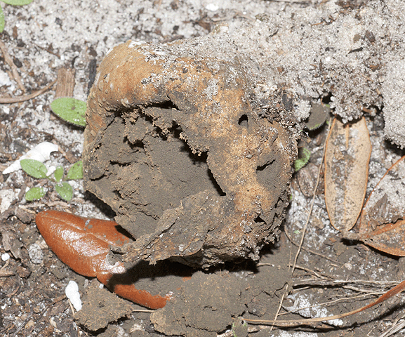
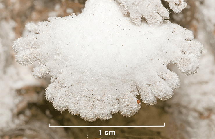
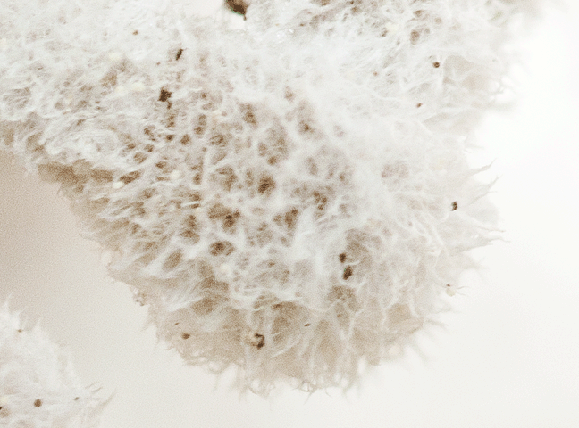
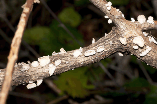
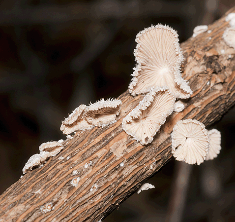
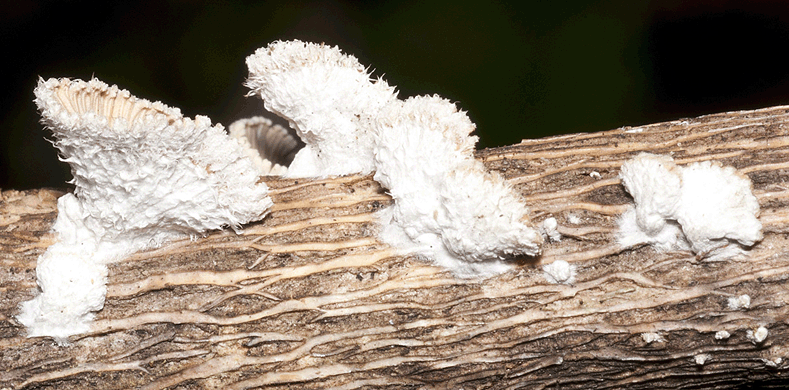
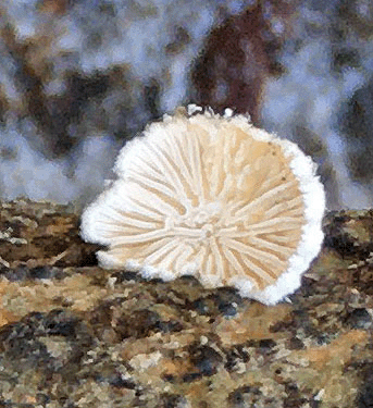
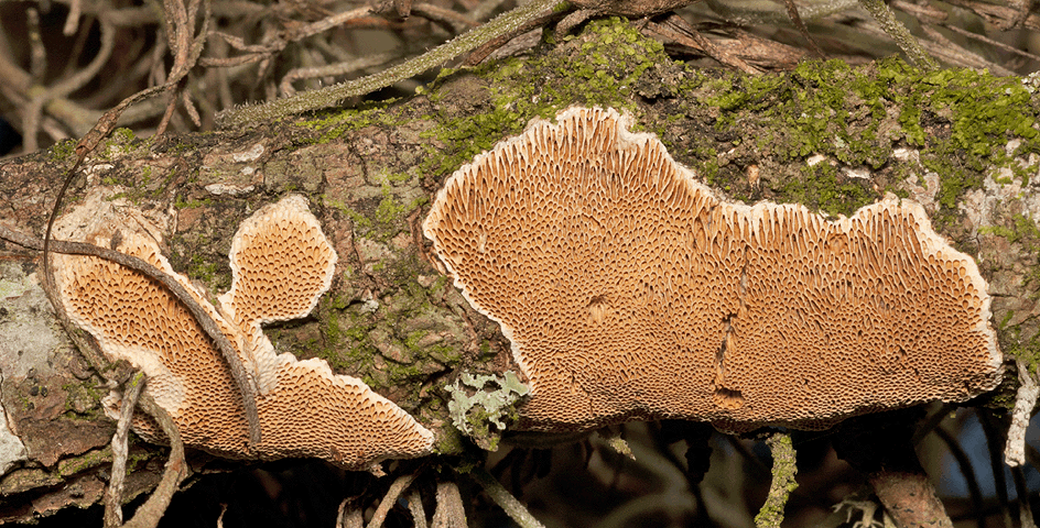
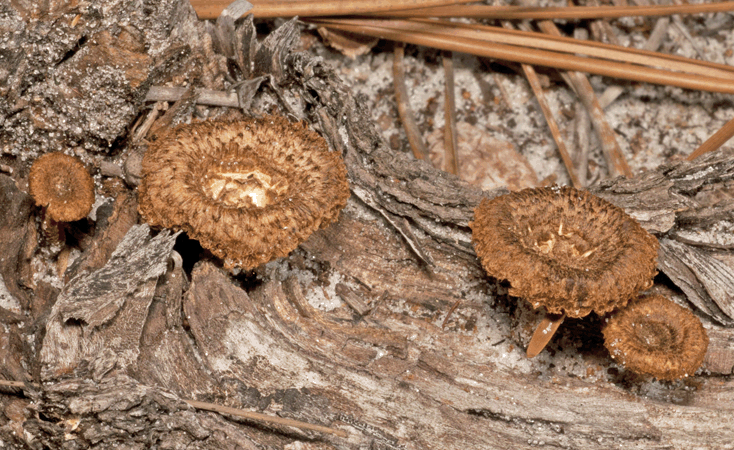
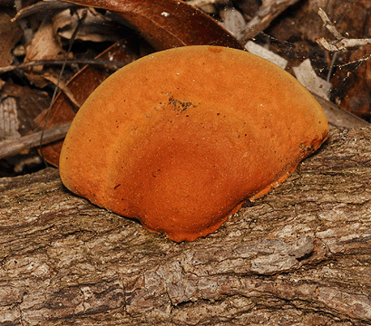
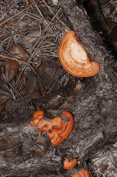
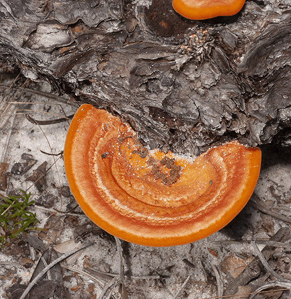
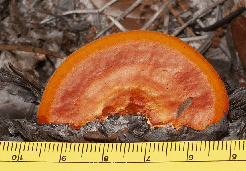
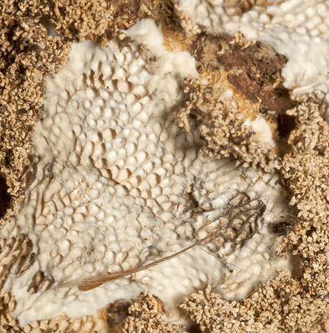
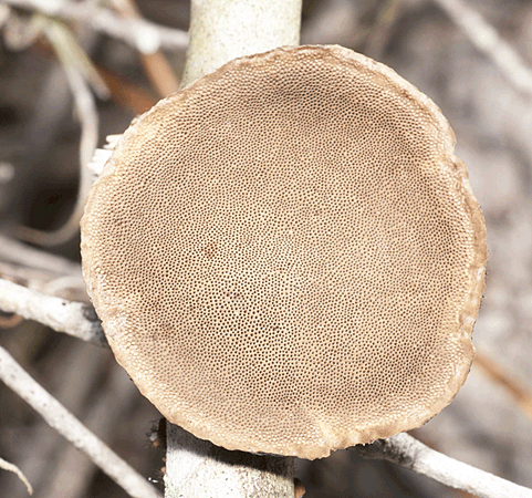 This fungus is a plant pathogen and is usually saprophytic, decaying dead wood tissues. The fungi shown here were growing on a branch that had been placed with other branches for a prescribed burn in the Smith Preserve.
 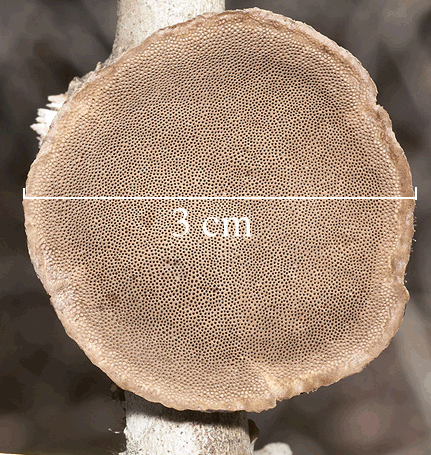 These images were sent to <mushroomobserver.org> for identification. The species was identified by Django Grootmyers (heelsplitter) on December 10, 2017.
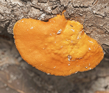 There are 5 species in the genus, separated by morphology, biogeography, and DNA sequence. Pycnoporus sangineus is found in warmer, tropical regions in North America, South America, and Asia.
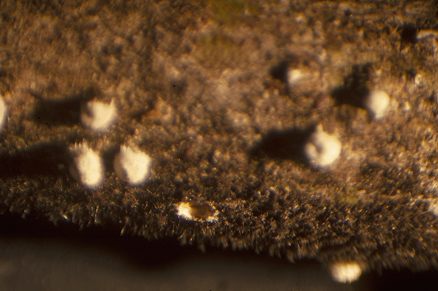
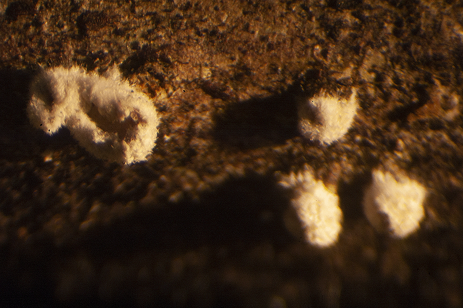
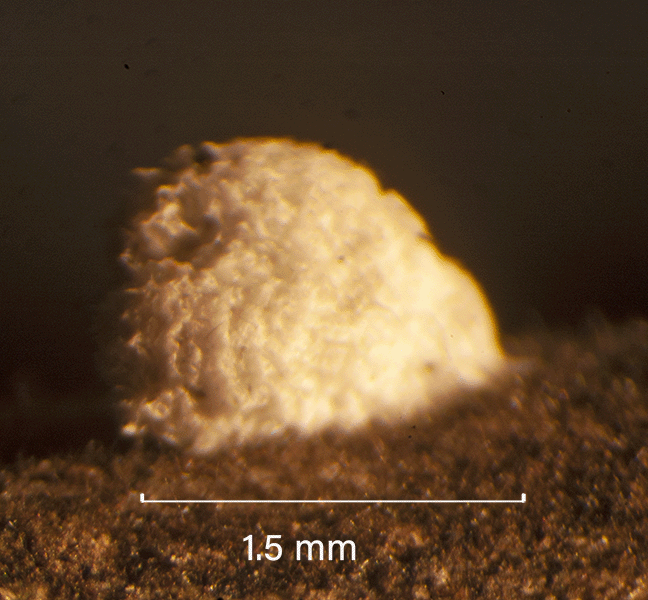
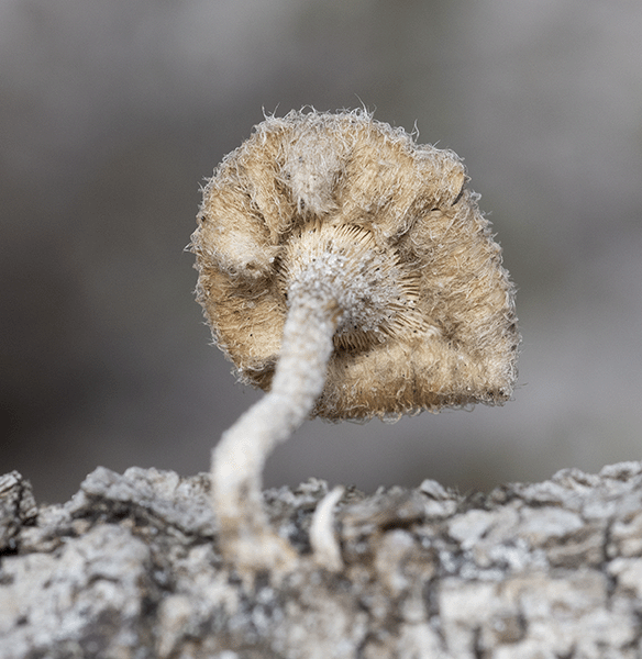
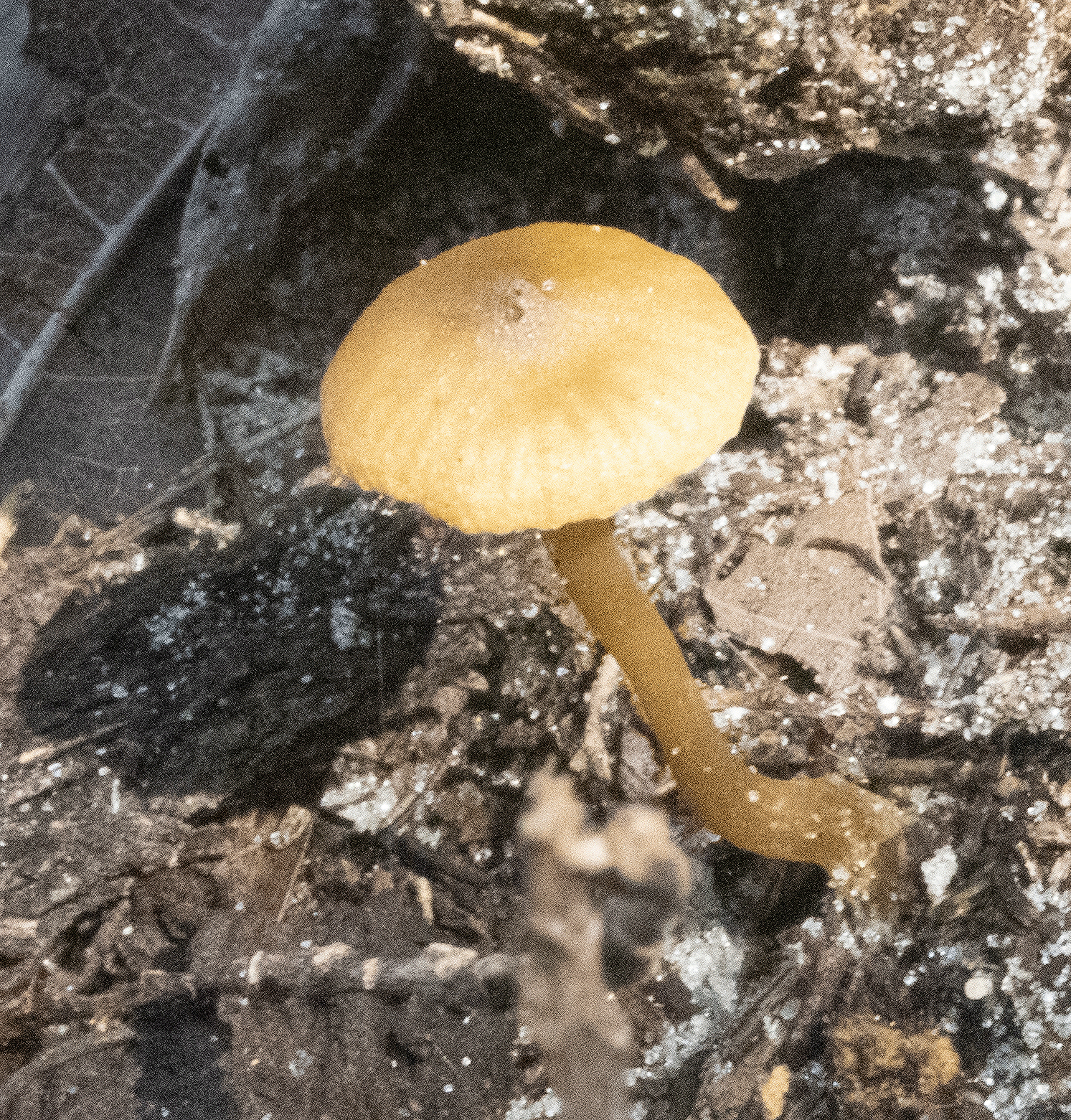
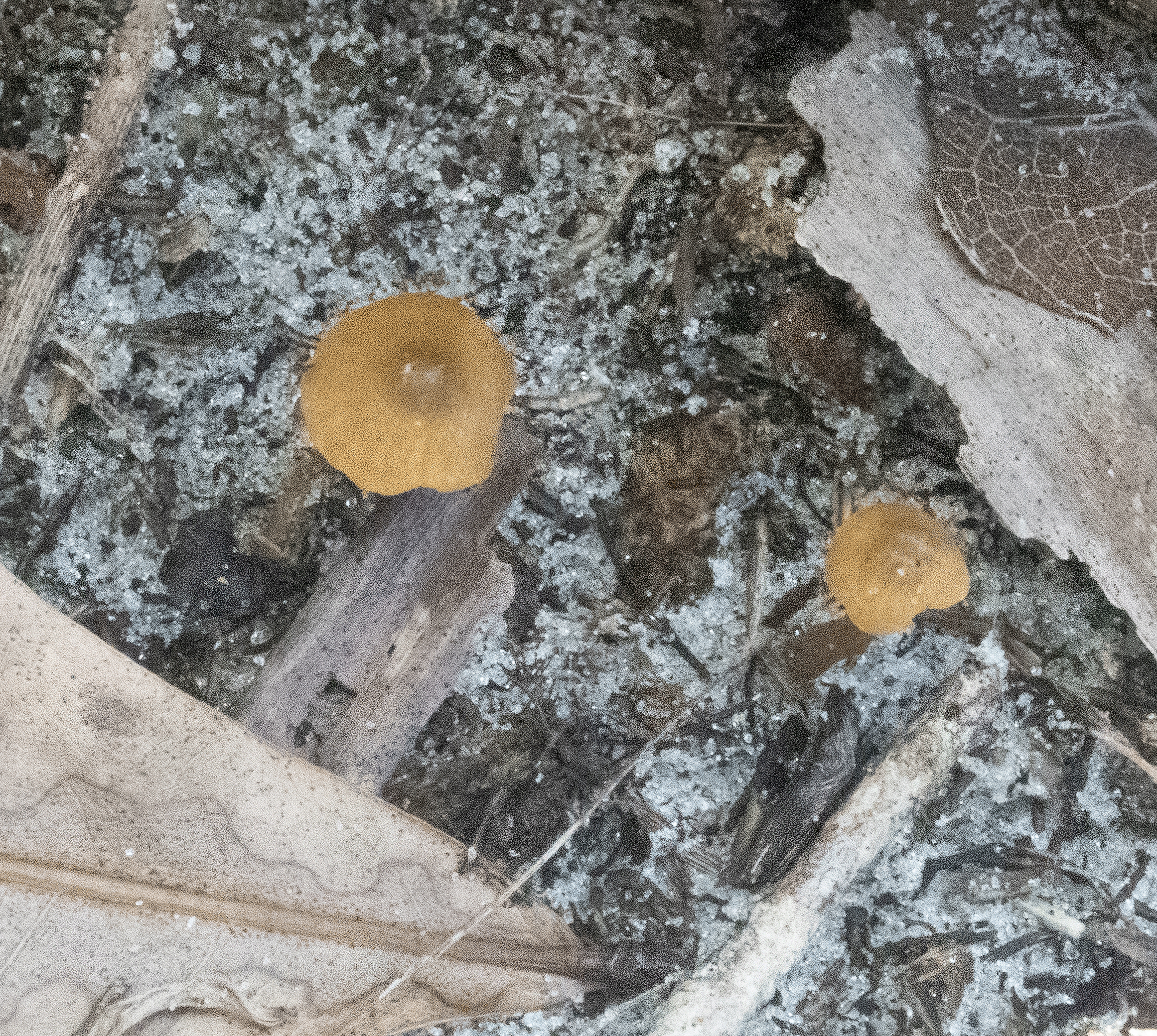
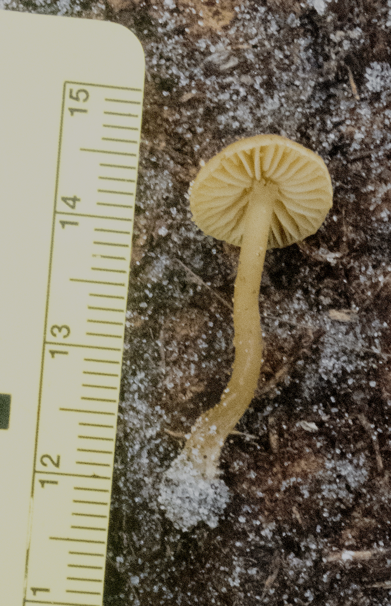
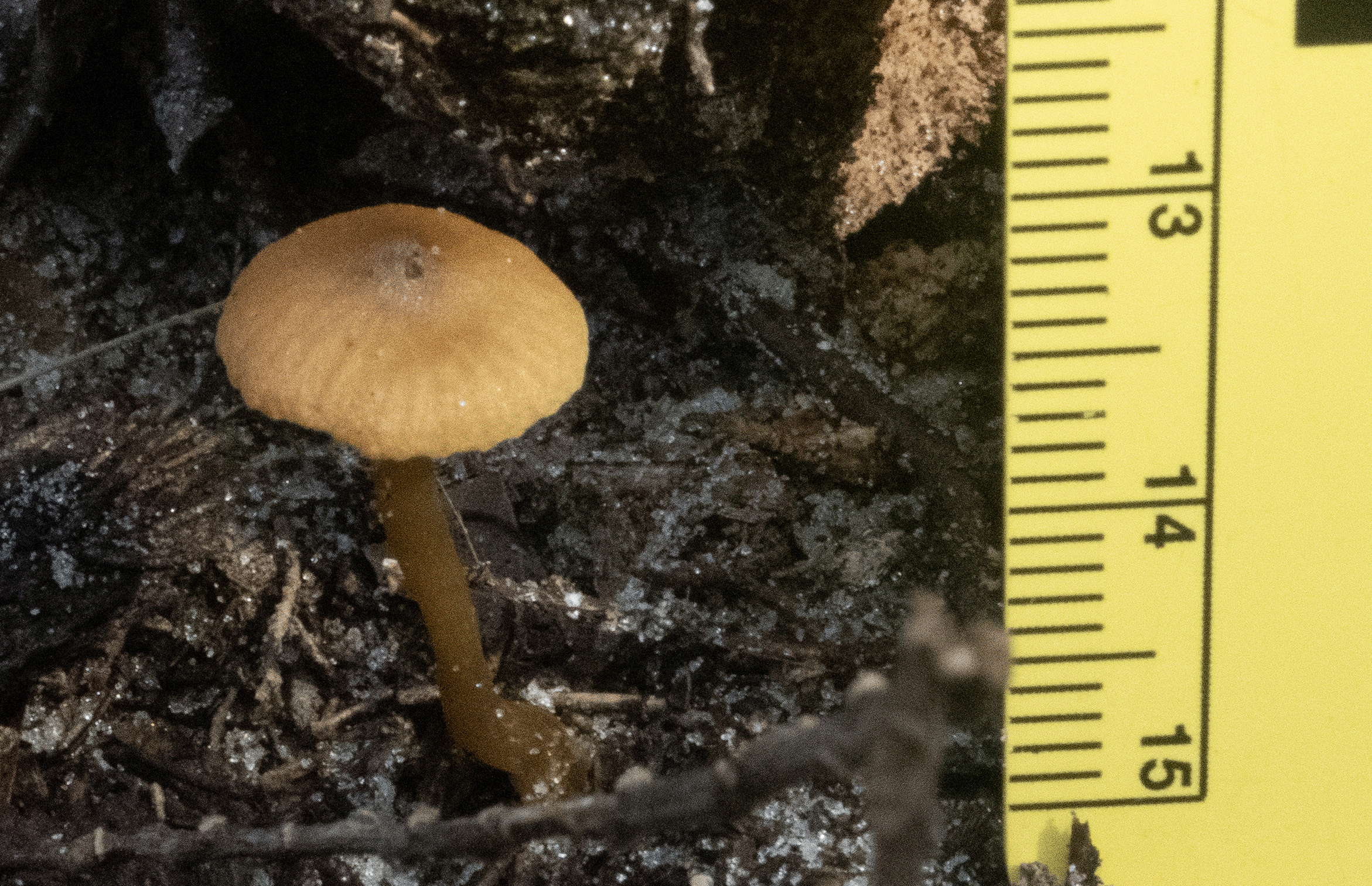
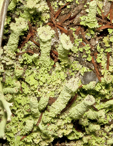
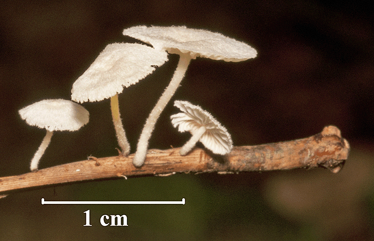
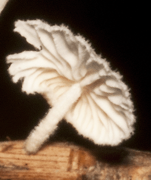
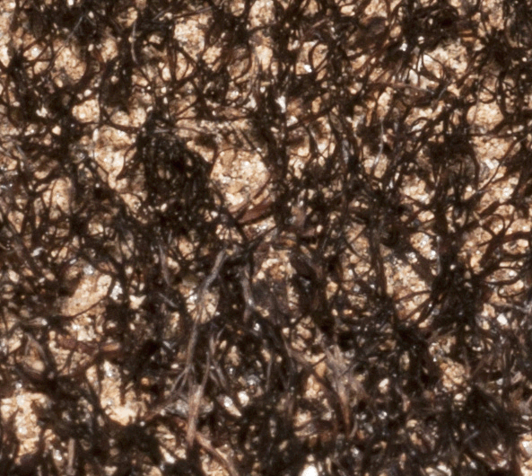
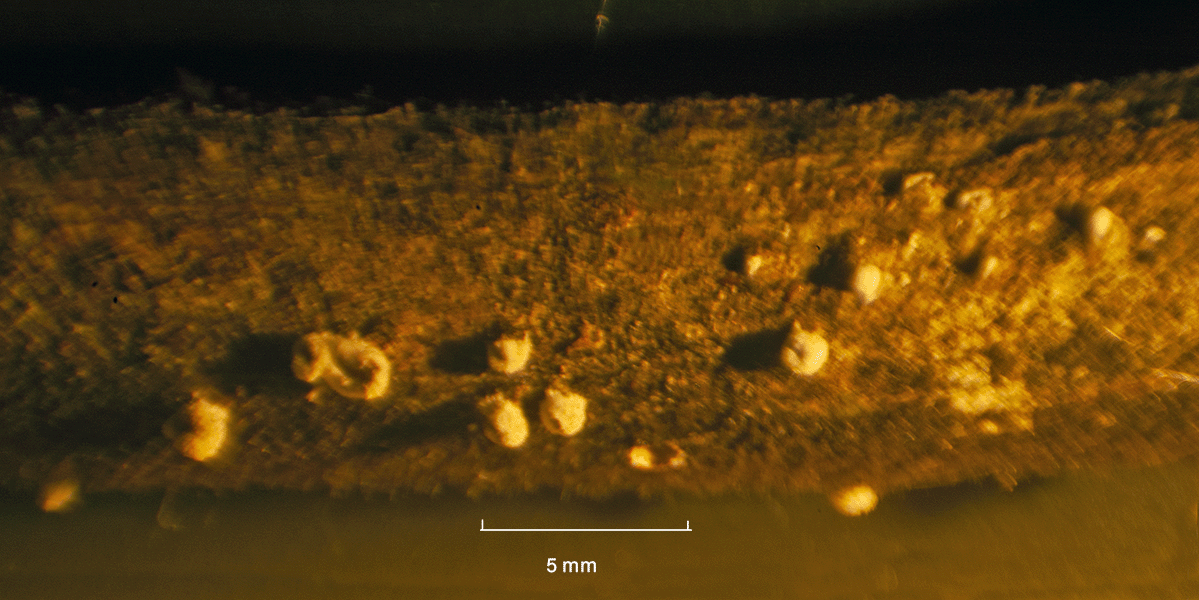 The photograph at right and all of those below were created through photomicrography. As can be seen, these fungi are very small. Many appear to be standing cylinders, with depressions at the top.